
Thanks to AI, the comprehensive #mesoSPIM-control documentation is finally here 🎇
mesospim.github.io/mesoSPIM-con...
09.03.2026 14:33
👍 3
🔁 1
💬 0
📌 0

@mesospim
Light-sheet microscopes for large samples. Open source hardware and software. Run by @nvladimus.bsky.social and @voigtvision.bsky.social Overview: www.mesospim.org Mailing list: http://eepurl.com/hPBRhj Forum: forum.image.sc/tag/mesospim
Thanks to AI, the comprehensive #mesoSPIM-control documentation is finally here 🎇
mesospim.github.io/mesoSPIM-con...

🚨Preprint alert🚨 Mapping of 3D collagen architecture with #tissueclearing, @mesospim.bsky.social #lightsheet #Microscopy, the Schmidt #multiimmersion microscope objective & #deeplearning in healthy tissue & wound healing by Wout Houbart & Thomas Naert & Co! www.biorxiv.org/content/10.6...

Schematic on how to correct field curvature along light-sheet propagation axis.
We're still exploring how to improve LSFM performance with "big" detection air objectives, where field curvature often limits achievable FOV. Here, Steven introduces curvedASLM, a method to dynamically correct field curvature (along one axis) in axially scanned setups www.biorxiv.org/content/10.6...


Fun to see photos from the @mesospim.bsky.social Symposium this last October.


The 10 Years of #mesoSPIM Symposium was a blast! 🔟🎂The excitement was palpable, and we were all inspired. Three days flew by fast! Thanks to all the attendees, speakers, organizing team, and our sponsors for making it such a memorable event! Let's shape the next 10y of microscopy together! 💪❤️🔬
📅 Start: 01.12.2025 (or on mutual agreement)
⏳ Application deadline: 31.10.2025
Curious? Learn more and apply here: t.uzh.ch/1U3
3/3
In this role, you will:
- Clear, stain, and image biological tissues
- Process and share imaging datasets
- Support and train platform users
We’re looking for candidates with lab experience, curiosity, and practical knowledge in tissue processing and fluorescence microscopy.
2/3

Job opportunity at the University of Zurich! 📢
We are looking for a 𝐋𝐚𝐛𝐨𝐫𝐚𝐭𝐨𝐫𝐲 𝐓𝐞𝐜𝐡𝐧𝐢𝐜𝐢𝐚𝐧 / 𝐑𝐞𝐬𝐞𝐚𝐫𝐜𝐡 𝐀𝐬𝐬𝐢𝐬𝐭𝐚𝐧𝐭 (𝟒𝟎%) to join our Microscopy Innovation Platform (MIP), an open-source initiative focused on advancing and making light-sheet and multiphoton microscopy accessible to researchers.
1/3
Both are experienced clinical pathologists who are master users of #mesoSPIM and developed their own way of staining and clearing the most challenging tissues in their clinical practice. Want to learn which method will work best with your samples? Join us at #mesoSPIM2025 t.uzh.ch/1Ss
3/3
Our invited speakers Dr. Anna Maria Reuss and Dr. Hei Ming Lai show astonishing results in this field with their new methods, #aDISCO and #INSIHGT, respectively, at the #mesoSPIM2025 symposium.
2/3

Image presenting two of the speakers for the 10 Years of mesoSPIM symposium. Background of the image is a microscopy picture showing blood vessels in magenta and glial cells in cyan on a black field. On the top left, the photo of the first speaker is contained in a white-rimmed circle. On the top right the text reads: "Hei Ming Lai, The Chinese University of Hong Kong, HK" followed by the talk title "Customizing mesoSPIM-based workflows for clinical 3D tissue Diagnostics". On the lower part of the image, the second speaker is presented in a specular manner. On the lower left, text reads “Anna Maria Reuss, University Hospital, Zurich, Switzerland”, followed by the talk title “aDISCO - A Broadly Applicable Method for 3D Microscopy of Archival Paraffin-Embedded Human Tissues”. On the lower left, the photo of the second speaker is contained in a white-rimmed circle. On the bottom right it reads “10 Years of mesoSPIM Symposium”
Is it possible to stain specific proteins and cell types in old formalin-fixed human tissues and make them transparent for light-sheet imaging and clinical 3D diagnostics?
🧵 1/3

Final program of the 10-year #mesoSPIM2025 Anniversary Symposium, October 13-15 in Zurich! Amazing lineup of speakers: biologists, physicists, developers, computer scientists and industry experts! Join us to learn, present, and connect! Registration until Sept. 15. www.adabd.uzh.ch/en/events/me...

📣We have opened our official User Forum: forum.image.sc/tag/mesospim
In this forum you can connect with 🔬developers and the image analysis experts. Ask your burning questions about the #mesoSPIM hardware, software, and image processing, or help the beginners to get started!
Tip: use project tags!

Image presenting one of the speakers for the 10 Years of mesoSPIM symposium. Background of the image is a microscopy picture showing blood vessels in magenta and glial cells in cyan on a black field. On the left, the photo of the speaker is contained in a white-rimmed circle. On the right the text reads: "Adam Glaser, Allen Institute, WA, USA" followed by the talk title "New technologies for mapping centimeter scale tissues with nanoscale resolution". On the bottom right it reads “10 Years of mesoSPIM Symposium”
How to image across scales from organs to synapses, unravel biological mechanisms and handle massive data?
Learn more from @adamkglaser.bsky.social, senior scientist at the Allen Institute and the lead developer of #ExA-SPIM technique, at the #mesoSPIM2025 Symposium, Oct 13-15.

mage presenting one of the speakers for the 10 Years of mesoSPIM symposium. Background of the image is a microscopy picture showing blood vessels in magenta and glial cells in cyan on a black field. On the left, the photo of the speaker is contained in a white-rimmed circle, on the right text reads: "Laura Baudis, University of Zurich, Switzerland" followed by the talk title "Probing dark matter and neutrinos with light sheet microscopy". On the bottom right reads "10 Years of mesoSPIM Symposium”
💥How can light-sheet help reveal dark matter?💥
Join Prof. Laura Baudis @lbaudis.bsky.social at #mesoSPIM Symposium, Oct 13–15 in Zurich or online, to see how light-sheet fluorescence microscopy 🔬 is used to image nuclear ☢️ recoil tracks to detect elusive particles!
Register now: t.uzh.ch/1Ss
What a pleasure to image such samples 🥰
#Deeplearning-based #CRISPR DNA integration and editing by Thomas Naert & Sören Lienkamp & Co with lots of @mesospim.bsky.social #lightsheet #Microscopy data of tadpoles (and some taken with the #Schmidt #multiimmersion objective prototype!) 🔬🐸 www.nature.com/articles/s41...

We’re excited to welcome @kevin-dean.bsky.social to the #mesoSPIM2025 Symposium! Explore his innovations in Multiscale Cleared-Tissue Axially Swept Light-Sheet Microscopy for imaging from organ to subcellular levels.
🗺️Oct 13–15 | Zurich & hybrid
Interested? Register now: t.uzh.ch/1Ss
Abstract Submission Deadline: TOMORROW, July 15-th 🎇

🎉 Call for Abstracts! 🎉
Abstract deadline approaching! 10 years of #mesoSPIM initiative – join us at the symposium for talks, demos & posters on microscopy, tissue clearing, and data analysis!
📆 October 13-15, 2025
🗺️ Zurich, Switzerland, but also online.
www.adabd.uzh.ch/en/events/me...
Speakers include @voigtvision.bsky.social @adamkglaser.bsky.social @christellelangevin.bsky.social @preibischs.bsky.social @jsdaniel02.bsky.social @nvladimus.bsky.social and many others! Poster design by @mgarbelli.bsky.social, cover image by Anna Maria Reuss.
Highlights:
🗣️ Keynotes & lightning talks
👩🔬 Maker Day for microscope builders
🤝 Meet devs, users & industry
Join us to shape the future of open-source microscopy!
#mesoSPIM2025 #openscience
Submit your work in any of these areas:
🧠 Bio/Neuro/Phys imaging
🧼 Tissue clearing techniques
🖥️ Image analysis tools (open or commercial)
🔧 Hardware and software (open or commercial)
Talks, posters & demos welcome!
Abstract submission: ema.uzh.ch/R7YRN

🎉 Call for Abstracts! 🎉
We’re celebrating 10 years of #mesoSPIM #opensource initiative – join us at the symposium for talks, demos & posters on open-source light-sheet microscopy, tissue clearing, and imaging!
Dates: October 13-15, 2025
Location: Zurich, Switzerland
Registration: ema.uzh.ch/R7YRN
Happy imaging!
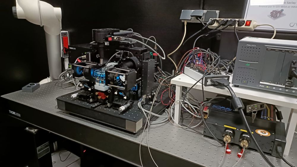
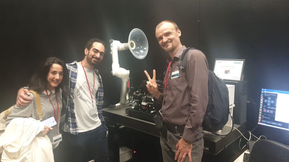
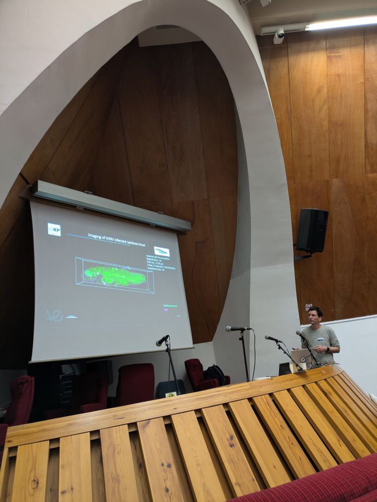
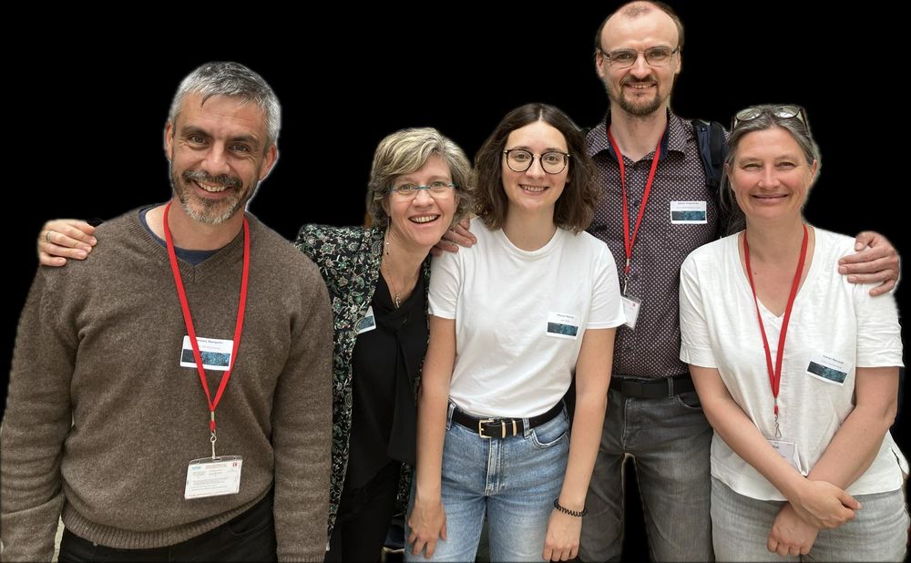
🔬 On June 6, we inaugurated the first #MesoSPIM in France at INRAE Jouy-en-Josas & ENS Paris-Saclay! A big thanks to @nvladimus.bsky.social , @mgarbelli.bsky.social ,Angeliki Vavladeli & all speakers and sponsors. Exciting step forward for #opensource imaging & interdisciplinary science! #microscopy

Did you know that Universite Paris Saclay has its own Benchtop #mesoSPIM? Interdisciplinary team lead by @christellelangevin.bsky.social has built their own setup, the first in France, and organized an inauguration symposium! Cleared baby sturgeon or trout? They have it! French bioimaging is strong!
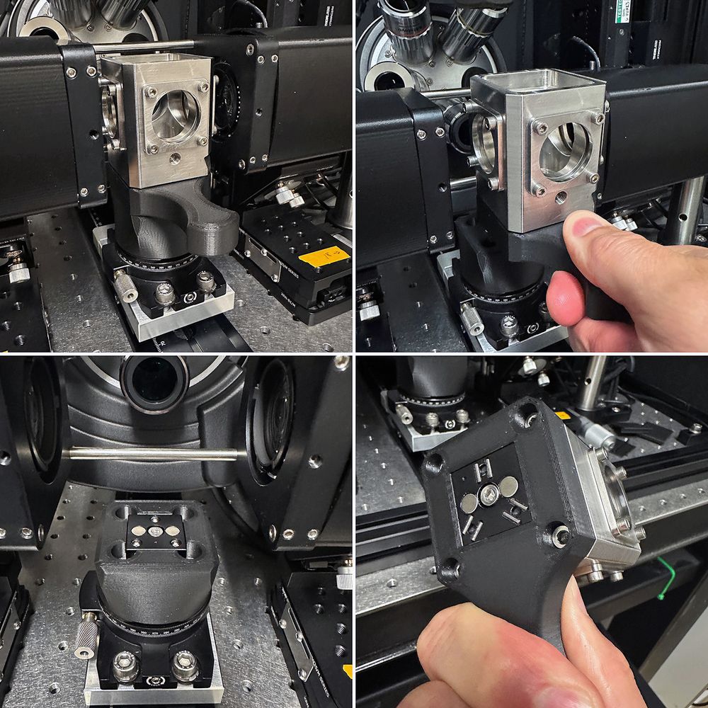
Stainless Imaging chamber for mesoSPIM with 3D printed kinematic mounts
New chamber mount design for our @mesospim.bsky.social - magnetic kinematic bases from @thorlabs.bsky.social for the win! 3D printed (for now) bases hold the chamber snugly for precise positioning between experiments. Fill port on the front allows for media addition without disturbing imaging.
Looking forward to the @mesospim.bsky.social #lightsheet #Microscopy anniversary symposium in October with @preibischs.bsky.social @adamkglaser.bsky.social @christellelangevin.bsky.social & many more! And to the next mesoSPIM decade! 🔬 Thanks @nvladimus.bsky.social & Co for organizing it!
