🌿Join us for the Webinar Series 2025 – Road to The 11th International Plant Biomechanics Conference 2026 🌍
📅 Webinar #1: Sept 25, 2025 | [Register: ugm.id/PBMweb1]
📅Webinar #2: Oct 9, 2025 | [Register: ugm.id/PBMweb2]
23.09.2025 16:12
👍 2
🔁 2
💬 0
📌 0
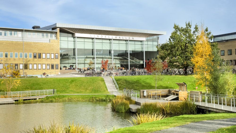
PhD position in plant science with a focus on cell adhesion
🧪🌾VACANCY - Do you have experience in cell and molecular biology and microscopy and are looking for a PhD?🔬
Join the multidisciplinary research group of @stephanevrg.bsky.social and study how plant cells adhere to each other.
Learn more here 👇 and apply!
www.umu.se/en/work-with...
21.05.2025 09:29
👍 7
🔁 7
💬 0
📌 2

Intracellular pathogens can form extensive hyphal structures. Here, the pathogen Phytophthora palmivora (magenta) produces invasive hyphae in a living epidermal cell of a Nicotiana root. The plant surrounds the invader by an 'extra-invasive hyphal membrane' (yellow)📸 @alexguyon.bsky.social
09.06.2025 16:31
👍 37
🔁 15
💬 0
📌 0

Epidermal cells acquire different cell shapes to enable their functions and maintain tissue integrity in plants. Meristematic cells differentiate and expand into diverse mature cell types, including jigsaw puzzle-shaped pavement cells (a), round stomatal guard cells (b), elongated epidermal cells in hypocotyls and the apical hook (c), and root epidermal cells with root hairs (d). The spatial distribution and arrangement of cell wall polysaccharides, such as cellulose microfibrils, xyloglucans, and pectins (e.g., homogalacturonan represent as HG), and the abundance of methylester groups on these components in differentiating cells regulate anisotropy during cell growth, enabling the acquisition of specific cell shapes. In addition, proteins like KATANIN and CLASP reorient dynamically the cortical microtubules. These cortical microtubules rearrangement is in response to mechanical cues, either self-generated (as in b, c) or from neighboring cells (a, c), leading to the resulting cell shape.
Attribution-NonCommercial-NoDerivatives 4.0 International

Auxin controls cell expansion by regulating cell wall biosynthesis and cell remodeling. Auxin promotes cell expansion by acidifying the cell wall and activating wall synthesis and loosening enzymes. Auxin efflux (PINs) and influx (AUX1) transporters establish concentration gradients in growing tissue. Auxin enters cells via influx transporters and activates the TRANSPORT INHIBITOR RESPONSE 1/AUXIN SIGNALING F-BOX PROTEINS-AUXIN/INDOLE ACETIC ACID (TIR1/AFB-Aux/IAA) nuclear signaling cascade, which regulates auxin-responsive genes, including AUXIN RESPONSE FACTORs (ARFs) and SMALL AUXIN UP RNAs (SAURs). Auxin activates the H+-ATPase proton pump through TRANSMEMBRANE KINASE 1 (TMK1), acidifying the cell wall and triggering loosening enzymes including PECTIN METHYLESTERASEs (PMEs), EXPANSINs, and XYLOGLUCAN:XYLOGLUCOSYL TRANSFERASEs (EXTs). Cellulose microfibrils are synthesized by the plasma membrane-bound cellulose synthase complex (CSC), with cortical microtubules guiding the exocytosis of this complex toward expanding cell edges. Auxin coordinates the reorientation of cortical microtubules and actin filaments to regulate the trafficking of cell wall polysaccharides to ensure proper cell wall expansion and specific cell shape acquisition.
Attribution-NonCommercial-NoDerivatives 4.0 International
🌱🧩 How do plant cells get their shape? 🧪
Check out the new #open-access #review exploring the mechanochemical duet between auxin & the cell wall in shaping diverse plant cell types from @srobertgroup.bsky.social's Lab.
🔗 doi.org/10.1111/ppl....
#PlantBiology #CellShape #Auxin #Biomechanics
30.05.2025 08:28
👍 27
🔁 11
💬 0
📌 1
⌛️#PlantBio2025 daily highlight: Advance Rates ending in few hours!🌿
Hurry and register soon for the best savings on the event you don’t want to miss!👉https://plantbiology.aspb.org/registration/.
#plantscience
10.06.2025 13:49
👍 6
🔁 6
💬 0
📌 0
📣Happening next Tuesday, June 17, 2025, at 5:00 pm Eastern Time. Join us!🌱
👉Free registration at plantae.org/plantaeprese....
#plantscience
10.06.2025 16:08
👍 9
🔁 4
💬 0
📌 0

IMPORTANT: The 11th International Conference on Plantbiomechanics has been scheduled to 2026. Please take note!
The venue and host continues to be: Universitas Gadjah Mada, Yogyakarta, Indonesia
14.02.2025 02:20
👍 3
🔁 4
💬 0
📌 1

Image text: Opportunity Alert! Now accepting applications for Summer Undergraduate Research Fellowships. Apply by February 7, 2025! Recipients receive: $6000 summer stipend, ASPB membership, $700 for materials, Travel support to Plant Biology 2026. Featuring ASPB logo on a plant themed background.
🚨 Opportunity Alert! Now accepting applications for 2025 Summer Undergraduate Research Fellowships (SURF).
Learn more and apply by February 7, 2025 👉 https://surf.aspb.org
21.01.2025 20:51
👍 19
🔁 12
💬 0
📌 2

🚨Hot off the press!
With Ibrahim Cheddadi, we tackled a key challenge: building a field theory of plant morphogenesis, based on fundamental balance laws and capturing cell wall remodelling and water dynamics in tissues🌿💧Check out our paper at www.sciencedirect.com/science/arti...
🧵👇
20.01.2025 10:18
👍 24
🔁 11
💬 5
📌 1

3D image of a flower meristem. Text overlay: Join the RESYDE project. 4 postdoc and 1 PhD. Research jobs and PhD studentships available at each institution are now open. Institutions are Humboldt-Universität zu Berlin, University of Cambridge, Umeå University and University of Sydney.
🚨JOB ALERT🚨
Seeking 4 Postdocs & 1 PhD to join €10 million multi-institution ERC Research #RESYDE project.
Help unravel the complex processes of symmetry breaking in plant development using flowers as a model system.
ℹ️ www2.hu-berlin.de/resyde/
Please share! #plantscijobs #PlantScience
16.01.2025 10:28
👍 25
🔁 20
💬 0
📌 1
The leptosporangium of Adiantum peruvianum shooting its spores, recorded with 50.000 fps
16.01.2025 15:03
👍 7
🔁 2
💬 0
📌 0
Good tips! Just editing a cover letter for @jxbotany.bsky.social now and they ask authors to answer these three questions:
1. What is the main question your manuscript seeks to answer?
2. How does your manuscript advance understanding of the topic?
3. Why is this work timely and important?
14.01.2025 20:29
👍 16
🔁 8
💬 0
📌 0

In just 5 month’s time we’ll be raising the curtain @debijloke in Ghent www.bijloke.be for Arabidopsis and much much more #ICAR2025 ! Registration is now OPEN, submit abstracts for oral presentations until March 17th, early bird registration runs until April 20th. More info on
www.icar2025.com !
15.01.2025 14:47
👍 14
🔁 7
💬 1
📌 0
The beauty (and mercilessness) of plant motion.
12.01.2025 20:29
👍 4
🔁 1
💬 0
📌 0
🔬☘️
Mechanical forces instruct division plane orientation of cambium stem cells during radial growth in Arabidopsis thaliana @currentbiology.bsky.social
doi.org/10.1016/j.cu...
Video: Cortical microtubules in cambium stem cells from hypocotyl longitudinal sections
26.11.2024 13:49
👍 26
🔁 9
💬 1
📌 0
![Figure 2: Homogalacturonan deficiency and rhamnogalacturonan-II
dimerization defects affect adhesion through largely separate pathways. (A-G)
Representative max intensity Z-projections of confocal stacks from 4-days-old darkgrown hypocotyl stained with propidium iodide from Col-0 (A), mur1-2 (B), qua2-1 (C),
mur1-2 qua2-1 (D), esmd1-1 (E), mur1-2 esmd1-1 (F) and qua2-1 esmd1-1 (G). (H)
Schematic representation of Boron-Rhamnogalacturonan-II (RG-II) dimers and
Calcium-Homogalacturonan (HG) “egg-box” crosslinking domains along the HG
backbone. (I) Quantification of the relative ruthenium red staining intensity (see
representative images and length quantification in figure S3). Boxplots summarize
three biological replicates, as represented by dots of different colors, and letters
describe the statistically significant differences between populations determined by
one-way ANOVA followed by Tukey’s HSD test (P < 0.05).[Scale bars, 30µm in images
(A-G).] See related content in figure S2 and S3.](https://cdn.bsky.app/img/feed_thumbnail/plain/did:plc:yizeaaccrv65tzu5cmxr7wv7/bafkreibyujocxmy7z2vea5qb2jw2vlm5zruz65dvzqkhublxtbok4hurii)
Figure 2: Homogalacturonan deficiency and rhamnogalacturonan-II
dimerization defects affect adhesion through largely separate pathways. (A-G)
Representative max intensity Z-projections of confocal stacks from 4-days-old darkgrown hypocotyl stained with propidium iodide from Col-0 (A), mur1-2 (B), qua2-1 (C),
mur1-2 qua2-1 (D), esmd1-1 (E), mur1-2 esmd1-1 (F) and qua2-1 esmd1-1 (G). (H)
Schematic representation of Boron-Rhamnogalacturonan-II (RG-II) dimers and
Calcium-Homogalacturonan (HG) “egg-box” crosslinking domains along the HG
backbone. (I) Quantification of the relative ruthenium red staining intensity (see
representative images and length quantification in figure S3). Boxplots summarize
three biological replicates, as represented by dots of different colors, and letters
describe the statistically significant differences between populations determined by
one-way ANOVA followed by Tukey’s HSD test (P < 0.05).[Scale bars, 30µm in images
(A-G).] See related content in figure S2 and S3.
🌱 New preprint from the Verger Lab @stephanevrg.bsky.social ! 🧪
📜 Imran Baba et al., 2024: doi.org/10.1101/2024...
🔬 Discover how RG-II crosslinking in the plant cell wall, regulated by Boron, is crucial for cell adhesion during growth.
✨ Bonus: RRQuant tool! (Details in thread 👇)
#PlantBiology
29.11.2024 14:54
👍 16
🔁 5
💬 1
📌 0

A postdoctoral position is available in the team of Gwyneth Ingram @rdplab.bsky.social as part of a collaborative project with our group @ibmp-cnrs.bsky.social
#plantscience
16.12.2024 17:10
👍 18
🔁 28
💬 0
📌 1

#PlantBio2025 in Milwaukee, Wisconsin (July 26-30), hosted by @aspbofficial.bsky.social features a plenary on #plantbiomechanics chaired by @erinsparks.bsky.social
plantbiology.aspb.org/plenary-symp...
Speakers: @geitmannlab.bsky.social, Naomi Nakayama,
@radinbio.bsky.social, Frank W. Telewski
10.01.2025 16:07
👍 7
🔁 1
💬 0
📌 2

Delighted to announce the 18th #FASEB Mechanisms of Plants Development will take place Aug 24-28th. ‼️ Register before June 29th → tinyurl.com/24fsjq6a
🌱 🎁 Vote for the conference image by Jan 31st
→ tinyurl.com/2872w6vm
The poster & speakers lineup will be announced in early Feb.
Spread the word!
08.01.2025 20:51
👍 58
🔁 49
💬 3
📌 1
If there was an award for plant-biomechanics-adjacent work, this one would definitely be a top candidate!
06.01.2025 15:27
👍 3
🔁 0
💬 0
📌 0

Job advertisement for image analysis expert
Job opportunity: Image Analysis Expert at @mcgill.ca, Montreal.
mcgill.wd3.myworkdayjobs.com/McGill_Caree...
RT!
#microscopy
#Imaging
#Imageanalysis
#Job
#JobOpportunity
#ResearchCareers
#OpenPositions
#BioImage
@royalmicrosoc.bsky.social
@vucellimaging.bsky.social
@microscopyirl.bsky.social
20.12.2024 17:14
👍 8
🔁 7
💬 0
📌 0

Job opportunity: CT Scan Research Assistant at @mcgill.ca, Montreal.
mcgill.wd3.myworkdayjobs.com/McGill_Caree...
RT!
#microscopy
#Imaging
#CTscan
#Radiology
#Job
#ResearchCareers
#OpenPositions
#BioImage
20.12.2024 17:21
👍 1
🔁 1
💬 0
📌 0
Plant Biomechanics friends and colleagues - I created a starter pack for our community. Feel free to let me know if you want to be removed from or added to the list!
go.bsky.app/D9oZmVH
22.12.2024 22:02
👍 19
🔁 11
💬 6
📌 0