Microscopy image of keratin (cyan) and desmosomes (magenta)
There are an extremely limited number of open slots for the Analytical and Quantitative Light Microscopy @aqlm.bsky.social course at @mblscience.bsky.social April 29-May 8 2026.
www.mbl.edu/education/ad...
Apply here for this amazing opportunity!
20.02.2026 17:52
👍 8
🔁 9
💬 0
📌 1

Figure showing the comparison between spinning disk (SD) confocal, SIM and ExM when imaging Drosophila cemtrioles.
Emma Burns, Anastasia Amoiroglou, Gregory Rogers, Nasser Rusan and colleagues develop an expansion microscopy protocol for Drosophila cultured cells and tissues, which they use to dissect centriole biology.
journals.biologists.com/jcs/article/...
22.01.2026 12:09
👍 17
🔁 9
💬 1
📌 0
Prime numbers 2-4999, animated with ggplot2+gganimate.
#Rstats code: gist.github.com/stephenturne...
20.01.2026 10:13
👍 67
🔁 21
💬 6
📌 2

Don't miss out. Apply to our 2026 Advanced Research Training Courses today!
15.01.2026 13:46
👍 18
🔁 16
💬 1
📌 1

Laboratory Engineer (Senior), in Advanced Imaging, 1.5.2026-31.12.2028
🚨 Job Alert! 🚨
We have an opening at our Microscopy Core Facility in Turku 🇫🇮.
If you love imaging 🔬 and helping scientists succeed, we want you! 👇
abo.rekrytointi.com/paikat/index...
#Microscopy #ScienceJobs #CoreFacility #Imaging
@turkubioscience.bsky.social
15.01.2026 14:04
👍 48
🔁 74
💬 0
📌 2


DC, Winter 2025
25.12.2025 22:05
👍 3
🔁 0
💬 0
📌 0

My lab in the NIH Intramural Program (Bethesda, MD) will be recruiting postdoctoral fellows over the next year with flexible start dates. We work on nanoscale cellular imaging of the plasma membrane and related organelles. Please reach out if you’re interested. www.training.nih.gov/jobs/pdp-053...
22.12.2025 14:57
👍 59
🔁 44
💬 1
📌 3
Addgene: Justin Taraska Lab Materials
Yes! Many of our plasmids are here www.addgene.org/Justin_Taras... and both NPY-GFP (exocytic vesicles) and clathrin light chain-GFP (endocytic sites) make excellent sub-diffraction biological targets.
22.12.2025 14:31
👍 2
🔁 0
💬 0
📌 0

Hoya bloom
30.06.2025 23:03
👍 6
🔁 0
💬 0
📌 0


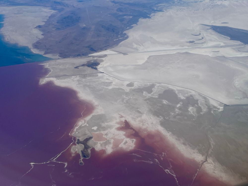
Salt Lake from the air
23.05.2025 01:30
👍 4
🔁 0
💬 0
📌 0

Metro
10.05.2025 14:35
👍 10
🔁 0
💬 0
📌 0
Japanese Garden, Powell’s Bookstore downtown, Forest Park, SE Division or SE Hawthorne St, Coava coffee, OHSU gondola.
07.04.2025 20:24
👍 3
🔁 0
💬 1
📌 0
31.03.2025 02:30
👍 15
🔁 0
💬 0
📌 0
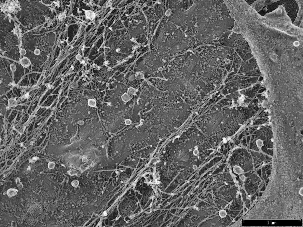
I am excited to share our first Pt replica EM images. It took us a little while, but now we have establish the unroofing, drying and Pt coating workflow 🎉 Great work by our postdoc Luis Wong Dilworth! The image below shows the cytosolic membrane leaflet of a fibroblast 👇
13.02.2025 14:33
👍 38
🔁 7
💬 2
📌 0

12.02.2025 15:39
👍 8
🔁 0
💬 0
📌 0
Super cool. Septin/actin rings maybe.
12.02.2025 15:14
👍 2
🔁 0
💬 1
📌 1

06.02.2025 14:31
👍 5
🔁 0
💬 0
📌 0

This is a scientific figure from the paper mentioned in the post. It shows fluorescence and electron microscopy images and associated schematics for using the ferritag to image clathrin coated pit proteins.
I'd like to draw your attention to this truly excellent paper from the Taraska lab on cryoET of plasma membrane associated proteins. Everyone who is thinking about probes in the cryoET space should also see what they could do with ferritag (fig 6) www.nature.com/articles/s41...
22.01.2025 16:40
👍 404
🔁 58
💬 11
📌 7

22.01.2025 02:53
👍 6
🔁 0
💬 0
📌 0
Not necessarily unhappy. Muscle cells have extensive flat clathrin lattices. Some cells stimulated with EGF also have them. Highly metastatic cells can accumulate them. They are used as adhesion and receptor organization sites.
14.01.2025 21:08
👍 3
🔁 0
💬 0
📌 0
Is the third image at the bottom surface of the cell? Those are likely large flat clathrin lattices that can accumulate in some cell types at the bottom of the cell.
14.01.2025 20:29
👍 3
🔁 0
💬 1
📌 0
02.01.2025 22:49
👍 6
🔁 0
💬 0
📌 0
CD44 and Ezrin restrict EGF receptor mobility to generate a novel spatial arrangement of cytoskeletal signaling modules driving bleb-based migration https://www.biorxiv.org/content/10.1101/2024.12.31.630838v1
01.01.2025 20:30
👍 2
🔁 1
💬 0
📌 0
It’s incredibly exciting when a completely different approach supports an unexpected finding from your group. Recently, we published that LDLR is captured with EGFR and FGFR1 at clathrin sites after EGF stimulation. I love science.
www.molbiolcell.org/doi/full/10....
bsky.app/profile/scie...
13.12.2024 18:21
👍 10
🔁 1
💬 0
📌 0