I am very proud of folks in my lab or who I know well who are applying to graduate school. They include 4 current postbacs, 2 summer students from Ghana, & 2 students I know well as a research sponsor or professor. All are so talented & most/all already have interviews scheduled! Look for them!
23.12.2025 17:10
👍 18
🔁 4
💬 0
📌 0


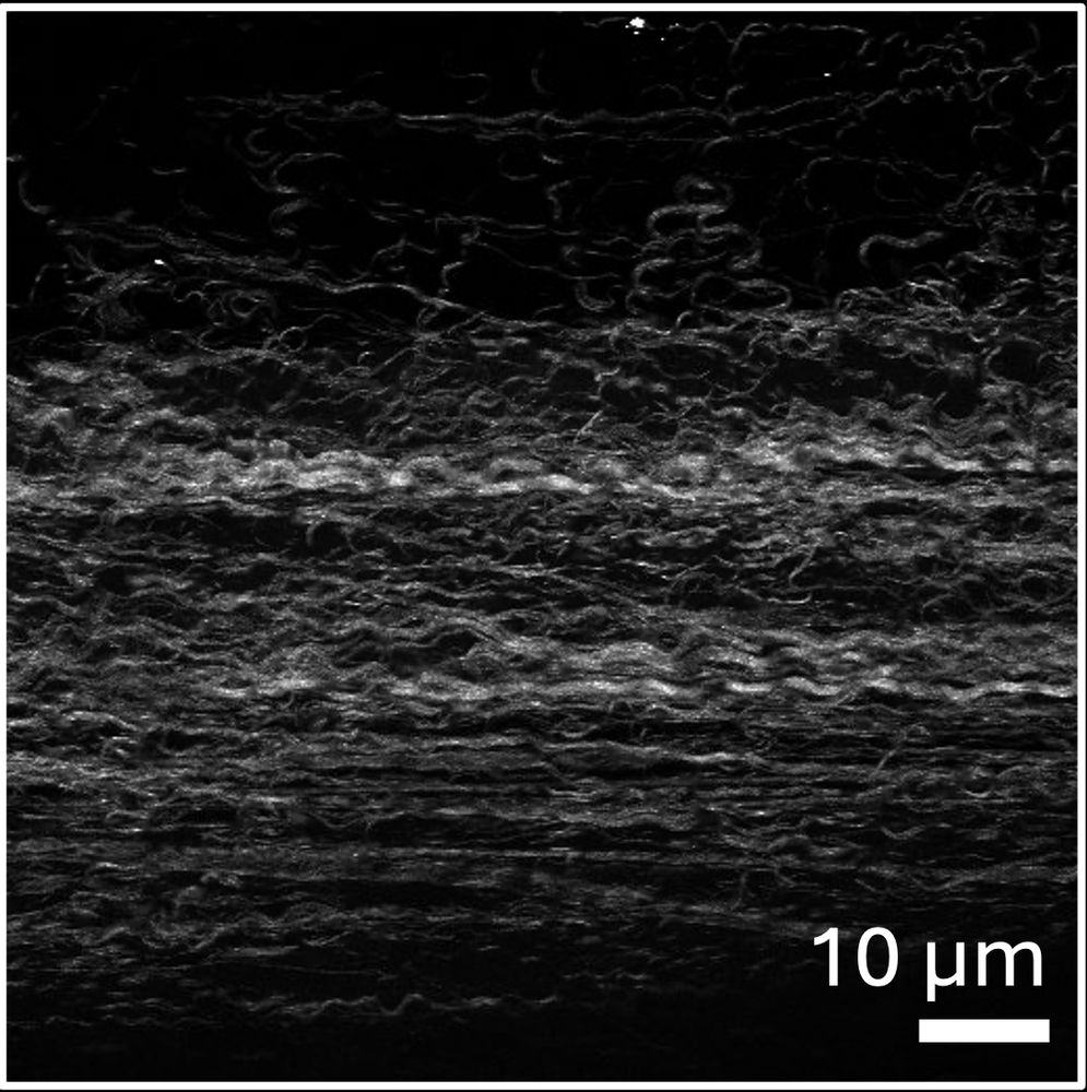
😃 In a new article published in J Biomech, Eekhoff et al. highlight the important function of collagen V in matrix assembly that has lasting effects into later ages, even though collagen V has little role in homeostatic maintenance of a mature tendon matrix.
👀 buff.ly/PZJ54C3
23.12.2025 20:12
👍 0
🔁 1
💬 0
📌 0
If you signed up to participate in SICB's mentor/mentee programs for the meeting in January. Check your email from Chair.SPDAC@sicb.org.
Please fill out the form link in the email before this Sunday, December 21!!
16.12.2025 22:01
👍 1
🔁 3
💬 1
📌 1

@tricytomeet.bsky.social continued with a lively poster session. I particularly liked the presentation by Naveen Chana, about work in Sid Shaw's IU lab on the plant augmin complex & microtubule regulation. She's now a PhD student in Joe Kieber's lab at UNC
trianglecytoskeleton.com/2025-meeting...
15.09.2025 15:53
👍 6
🔁 2
💬 0
📌 1

Microscopic picture of Drosophila Sperm (green, yellow, orange, red) and Muscle cells (teal, blue). Depth coded.
Happy #FluorescenceFriday !🌈
Today, sperm cells & developing gametes (Dapi, 🟢🟡🟠🔴) and testis muscles (Phalloidin (🔵🟢) in/on a #Drosophila non-melanogaster (guess the species and you’re the 🐐)
#cellbio #devbio #science #sciart #microscopy
Scope: Zeiss Airyscan 980❤️@zeiss-microscopy.bsky.social
06.06.2025 14:08
👍 70
🔁 12
💬 2
📌 1

Six fish arrayed diagonally over the top of a rainbow pride flag. At the top left on the red stripe is a northern redbelly dace (Chrosomus eos), on orange is a male rainbow darter (Etheostoma caeruleum), a shorthead redhorse (Moxostoma macrolepidotum) on the yellow stripe, a northern pike (Esox lucius) for the green stripe, an orangespotted sunfish (Lepomis humilus) on the blue strip and a goldeye (Hiodon alosoides) on the purple stripe.
Happy Pride! The diversity of life, in all its forms, is pretty rad!
(Side note: there's not a whole lot of purple fish, so the pink/silver goldeye is the best I got)
#SundayFishSketch #Pride
08.06.2025 16:23
👍 94
🔁 24
💬 1
📌 0

An illustration of a Rainbow Trout, but instead of the realistic coloring on the body, the colors make a rainbow in honor of Pride month.
I've been camping almost every weekend as of late so haven't been able to post #SundayFishSketch, but here is an edited version of my Rainbow Trout illustration with altered coloring in honor of #PrideMonth.
07.06.2025 11:39
👍 96
🔁 23
💬 1
📌 0

Hi #FluorescenceFriday community! 🌈
Fly testes are unexpectedly stunning 😀
Here’s a testis from a non-melanogaster Drosophila species:
🟠🟡 DNA: Dapi
🔵🟢 Muscles: Phalloidin
LUT: @kwolbachia.bsky.social
Scope: @nat-prunet.bsky.social 's core
#sciaart #microcosm #closeup #microscopy #cellbio
04.04.2025 17:45
👍 59
🔁 8
💬 4
📌 0

My single favourite wildlife photo from our trip to Galápagos has to be this marine iguana sneezing out salt - such an elegant and glamorous adaptation to their unusual lifestyle!
12.03.2025 17:00
👍 106
🔁 11
💬 1
📌 1
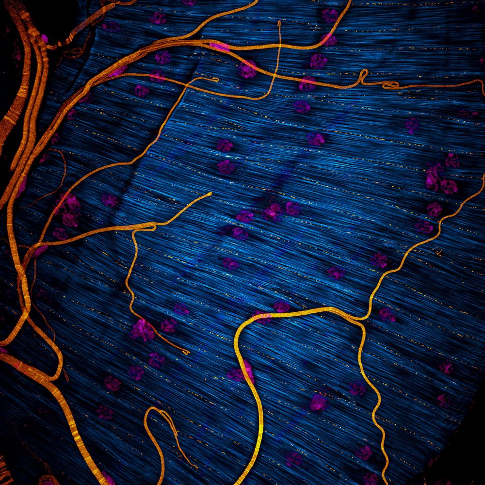
2/4 I chose this picture because I am interested in an old question:
How do fly testes obtain their specific shape?
In my previous research I learned that muscle precursors shape it, using collective cell migration and alignement to achieve the pattern visible in this picture.
11.03.2025 12:03
👍 33
🔁 7
💬 6
📌 1

Vote for your favourite image - FocalPlane
Vote for your favourite image - News
1/4 Super excited to take part in my first big microscopy competition. My picture is the one with the Drosophila hydei testis musculature (Invisible Architects, #12).
focalplane.biologists.com/2025/03/11/v...
All pictures in this list are fantastic, so, please go vote! 😊🔬
#science #microscopy
11.03.2025 12:03
👍 49
🔁 11
💬 2
📌 3

Flier announcing Jonathan's PhD defense seminar. Photo of salamander in the corner. Zoom link upon request.
Among all the bad, there is some good. Happy to announce that I will defending my PhD in two weeks. March 24 at 2pm EST. Message me if you'd like the Zoom link.
11.03.2025 01:07
👍 63
🔁 5
💬 7
📌 1
Post the amazing science things you have done with federal funding.
28.01.2025 20:51
👍 1553
🔁 602
💬 170
📌 317
Ooohhh-that's our paper! Check it out! @noahgurley.bsky.social @emily-mcparland.bsky.social
04.01.2025 01:17
👍 35
🔁 9
💬 0
📌 0
I'm actually the student rep for Division of Vertebrate Morphology 😊 way ahead of you there
04.01.2025 17:58
👍 2
🔁 0
💬 1
📌 0
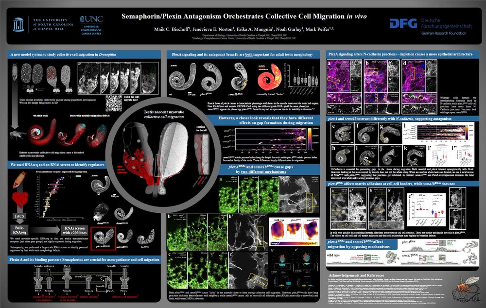
#CellBio2024 If you like collective cell migration, organogenesis, or very cool movies check out @maikbischoff.bsky.social's poster this morning (Tuesday, Dec 17 11:15 AM) Semaphorin/Plexin Antagonism regulates Collective Cell Migration and Gap Closure in vivo [P2780- Board B397]
17.12.2024 16:00
👍 30
🔁 9
💬 1
📌 0

The head of a dark colored shortnose gar in a well planted aquarium tank.

Three gar, one shortnose and two longnose, in a well planted aquarium tank with other fish in the background.

Two gar fossils in a museum display case.
Visited Chicago just in time for #Garweek and #fishmas! Saw live Shortnose & Longnose gar at Shedd Aquarium, then fossils at the Field Museum. The living gar's DNA is very similar to their ancient relatives!
16.12.2024 23:05
👍 26
🔁 7
💬 2
📌 0
Our work on how cell junction-cytoskeletal linkage is regulated is out. Great work by @emily-mcparland.bsky.social, @noahgurley.bsky.social & others. Thanks to @jcellsci.bsky.social for straightforward review & for open access via a Read & Publish agreement 🧪
journals.biologists.com/jcs/article/...
15.12.2024 13:40
👍 107
🔁 27
💬 4
📌 1

Picture of Emily and the intro to the article in the link

Picture of Noah and one section of the article in the link
Hey #CellBio2024 - Another amazing thing about publishing in @jcellsci.bsky.social are their "First Person" interviews with the scientists who actually did the work. Here's a new one featuring lab alums @emily-mcparland.bsky.social & @noahgurley.bsky.social
journals.biologists.com/jcs/article/...
15.12.2024 14:53
👍 24
🔁 7
💬 0
📌 0

Noah Gurley

Emily McParland
Read more about this research in our ‘First person’ interview with Emily McParland and Noah Gurley: journals.biologists.com/jcs/article/...
17.12.2024 13:06
👍 2
🔁 1
💬 0
📌 0

Immunofluorescence images of embryos showing that CnoRA1 domain is important for dorsal closure, head involution and epidermal integrity.
Wild-type (WT) embryo. Some cells are rounded up to divide (white arrows), but they rapidly return to columnar architecture. (B) cnoΔRA1. Groups of cells near the ventral midline fail to resume columnar architecture (red arrows). (C–E) Stage 11. (C) Wild-type embryo. (D,E) cnoΔRA1. Failure to resume columnar architecture becomes more apparent (red arrows). (F–H) Stage 14. (F) Ventral view of a wild-type embryo. Dorsal closure is completed (white arrow), head involution is underway (yellow arrow) and segmental grooves are shallow (green arrowheads). (G) cnoΔRA1. Dorsal closure failed, exposing underlying tissues (red arrow). There are gaps in the head epidermis (yellow arrows) and deep segmental grooves remain (green arrowheads). (H) Ventral view, cnoΔRA1. Holes in the epidermis (red arrows) and deep segmental grooves (green arrowheads) are observed. (I–J) Closeups, wild-type stage 10 (I–I″) and 11 (J). Dividing cells (green arrows) and forming tracheal pits (cyan arrows) are observed. Arm, Baz and Cno remain enriched at AJs. (K–K‴) Stage 10/11 cnoΔRA1 mutant. Cells that retained columnar architecture retain junctional Arm, Baz and CnoΔRA1 (yellow arrows). However, in some cells AJs are fragmented (red arrows) and in less epithelial regions Arm, Baz and Cno are strongly reduced (cyan arrows).
@emily-mcparland.bsky.social, @noahgurley.bsky.social, Kevin Slep, @peiferlabunc.bsky.social and colleagues dissect the differential roles of the dual Ras-association domains of Drosophila Canoe in linking cell junctions to the cytoskeleton.
journals.biologists.com/jcs/article/...
17.12.2024 13:06
👍 14
🔁 6
💬 1
📌 0

This is the face of a shark! I've stained the mineralised tissues to show that their teeth and scales are both made of the same enamel-like material 🦈 🦷 🧪
11.12.2024 10:01
👍 309
🔁 72
💬 9
📌 11
A renewed reminder to anyone applying for a PhD or postdoc position in a lab--indicate your interest in the email/cover letter by talking a bit about that lab's work and why it would be a good fit. I answer any query that includes this information but do not answer those that do not.
05.12.2024 13:55
👍 133
🔁 29
💬 6
📌 4
Noah is so cool!
05.12.2024 16:04
👍 2
🔁 0
💬 0
📌 0
An amazing overview of our work that is now in press at @jcellsci.bsky.social
Super lucky to work with this amazing team of scientists.
05.12.2024 16:04
👍 12
🔁 1
💬 0
📌 0

Title authors and abstract from the link
In work now in press @jcellsci.bsky.social , we explored this, combining bioinformatic, biochemical, genetic and cell biological tools. This was powered by a remarkable team of postbac scholars including @emily-mcparland.bsky.social
journals.biologists.com/jcs/article/...
30.11.2024 14:32
👍 34
🔁 10
💬 2
📌 0

A cicada emerges from its husk, all pink and purple and shiny
Congratulations. You’ve survived until Wednesday. Here, have a cicada, as a treat.
#inverts #invertebrate 🐡
20.11.2024 14:33
👍 1477
🔁 100
💬 62
📌 8
Hello Blue Sky! SPDAC is planning a bunch of stuff for SICB this year! Stay tuned 👀
17.11.2024 23:52
👍 4
🔁 1
💬 0
📌 0